Staged Approach of Acute Limb Ischaemia on the Background of Popliteal Artery Aneurysm
| Available Online: | July, 2025 |
| Page: | 85–89 |
Author for correspondence:
Emmanouil Barmparessos MD, MSc
Vascular Surgery Department, University Hospital of Patras
Rio, 26 504, Greece
Tel: +30 6946519511
E-mail: mparmpar@ac.upatras.gr
ISSN 2732-7175 / 2025 Hellenic Society of Vascular and Endovascular Surgery
Published by Rotonda Publications
All rights reserved. https://www.heljves.com
Full Text
Full Text
Abstract:
Patients with Popliteal Artery Aneyrysm (PAA) may present, quite often, with Acute Limb Ischaemia (ALI) and despite treatment the risk of amputation remains high due to compromised run-off vessels. Sufficient evidence regarding the techniques to improve the run-off vessels in acute settings remains scarce with literature being inconclusive so far. Herein, we report a case of ALI on the background of PAA which was treated in a stepwise approach. The PAA was initially excluded by open repair through a posterior approach followed by run-off vessel restoration through a combination of endovascular techniques. Procedure was successful and highlights the importance of research of the new available techniques through a multidisciplinary team.
Keywords: Acute Limb Ischaemia, Popliteal Artery Aneurysm
Introduction
A significant proportion of patients with Popliteal Artery Aneurysm (PAA) may present acutely with limb ischaemia. Given the acute settings, the compromised run-off vessels and the absence of robust data from well-controlled studies to support the decision making, the current pathology remains demanding. Herein, we report a case of Acute Limb Ischaemia on the background of PAA (ALI-PAA) which was treated in a staged manner by combining open surgery and endovascular techniques. Informed consent was obtained from the patient before publishing images and history; therefore, approval by the institutional review board was waived.
Case presentation
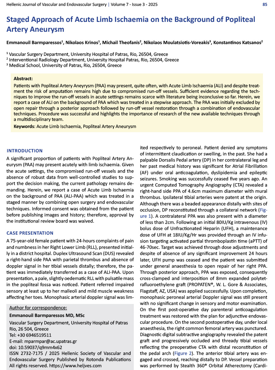
A 75-year-old female patient with 24-hours complaints of pain and numbness in her Right Lower Limb (RLL), presented initially in a district hospital. Duplex Ultrasound Scan (DUS) revealed a right-hand side PAA with parietal thrombus and absence of doppler signal in her tibial vessels distally; therefore, the patient was immediately transferred as a case of ALI-PAA. Upon presentation, a pale, slightly oedematic RLL with pulsatile mass in the popliteal fossa was noticed. Patient referred impaired sensory at least up to her malleoli and mild muscle weakness affecting her toes. Monophasic arterial doppler signal was limited respectively to peroneal. Patient denied any symptoms of intermittent claudication or swelling, in the past. She had a palpable Dorsalis Pedal artery (DP) in her contralateral leg and her past medical history was significant for Atrial Fibrillation (AF) under oral anticoagulation, dyslipidemia and epileptic seizures. Smoking was successfully ceased five years ago. An urgent Computed Tomography Angiography (CTA) revealed a right-hand side PPA of 4.6cm maximum diameter with mural thrombus. Ipsilateral tibial arteries were patent at the origin. Although there was a beaded appearance distally with sites of occlusion, DP reconstituted through a collateral network (Figure 1). A contralateral PPA was also present with a diameter of less than 2cm. Following an initial 80IU/Kg intravenous (IV) bolus dose of Unfractionated Heparin (UFH), a maintenance dose of UFH at 18IU/Kg/Hr was provided through an IV infusion targeting activated partial thromboplastin time (aPTT) of 46-70sec. Target was achieved through dose adjustments and despite of absence of any significant improvement 24 hours later, UFH pump was ceased and the patient was submitted under general anaesthesia to open repair of her right PPA. Through posterior approach, PPA was exposed, consequently cross-clamped and interposition of 8mm expanded polytetrafluoroethylene graft (PROPATEN®, W. L. Gore & Associates, Flagstaff, AZ, USA) was applied successfully. Upon completion, monophasic peroneal arterial Doppler signal was still present with no significant change in sensory and motor examination. On the first post-operative day parenteral anticoagulation treatment was restored with the plan for adjunctive endovascular procedure. On the second postoperative day, under local anaesthesia, the right common femoral artery was punctured. Diagnostic digital subtractive angiography revealed the patent graft and progressively occluded and thready tibial vessels reflecting the preoperative CTA with distal reconstitution of the pedal arch (Figure 2). The anterior tibial artery was engaged and crossed, reaching distally to DP. Vessel preparation was performed by Stealth 360® Orbital Atherectomy (Cardiovascular Systems Inc., St. Paul, MN,) through the 1.25mm Micro Crown. Subsequently treatment was applied through balloon angioplasty by 3x22mm Ultraverse 014 (BD Interventional, 3rd Street Tempe, AZ, USA) as well as 2.5x100mm CoyoteTM, (Boston Scientific, Marlborough, MA, USA) (Figure 3a,3b,3c). Eventually, as the improvement of the inflow was succeeded by balloon angioplasty (6x40mm Passeo-18, Biotronik AG, Bülach, Switzerland) aiming at a significant local stenosis at the level of Hunter’s canal (Figure 3d), the completion angiography revealed brisk flow to the DP (Figure 4). Clinical examination was significant for a palpable right DP. Although numbness and motor deficit of toes persisted there was no evidence supporting compartment syndrome. Given the history of AF, oral anticoagulation was reinstituted with a 3-month addition of Clopidogrel 75mg. The patient gradually convalesced uneventfully and regained complete function. At 3 months follow-up, patient denied any limb-relating symptoms, the graft was patent on Duplex Scan with palpable DP. Surveillance at 6months interval will continue, monitoring her contralateral PPA.
Discussion
PAA has been reported as a rare disease, albeit the most common peripheral aneurysm. In almost 30 percent of the cases the presentation may be acute with symptoms of thrombosis and despite treatment the risk of amputation is reported as high as 14%1. The reason of the acute limb ischaemia may be the PAA thrombosis or distal embolisation or combination of them. Given the extensive mural PAA thrombus, the diminished collateral and the occupied tibial arteries through chronic distal embolization, the caused ALI may be severe and very challenging to treat specially in acute and out-of-hours settings2. In our case a narrow lumen inside the PAA of the affected limb was preserved upon presentation. However, the distal tibial arteries had been progressively packed with emboli in combination with multifocal calcified stenoses which in fact “helped” to preserve the pedal arch. A recent Vascunet report, showed that there is significant variability in the approach of PAA3 calling the physicians to reconsider the endovascular repair (ER). Beuschel et al. conducted a systematic review and metanalysis of treatment and natural history of PAAs showing that open repair (OR) is more durable than ER4. Based on that study the Society of Vascular Surgery (SVS) published guidelines5 dedicated to PAAs were although they do not recommend against ER, as the European Society of Vascular Surgery did with the guidelines on ALI6, they recommend that the management of ALI-PAA should be based on the severity of ALI on presentation. More specifically, SVS recommend thrombolysis or pharmacomechanical intervention (PMI) to improve the runoff vessels for patients with mild or moderate ALI (Rutherford I and IIa) and prompt OR or ER with adjunctive thromboembolectomy (TEE) or PMI for severe ALI (Rutherford IIb). There is a controversy in literature regarding the preoperative thrombolysis (PT) with Kropman et al reporting no significant benefit on amputation rate by analysing data of 895 ALI-PAA cases1 and on the other hand Ravn et al. reported significantly better results for PT on 235 ALI-PAA patients; however both of these studies lack of data regarding the severity of ischaemia. More recent studies focusing on the intraoperative thrombolysis (IT) during OR in ALI-PAA showed improved outcomes7,8. The complexity of the ALI-PAA management is reflected in the compromised run-off. In our case, a potential tibial TEE through medial approach would cause more damage than outflow restoration given the beaded, calcified appearance as well as the size of the vessels. On the other hand, given the thrombotic burden of the PAA, any endovascular intervention prior to aneurysm exclusion would pose the risk of further embolic events7. Therefore, we chose the posterior approach to exclude the aneurysm with the least insult and on a second stage improve the outflow through endovascular approach. Any potential delay between the stages would have risked the bypass, hence a good collaboration in a multidisciplinary team is crucial. As far as we are concerned, this is the first report in English literature of a staged approach to treat ALI-PAA, where initial OR excluded the PAA and subsequently endovascular techniques safely restored the run-off.
Conclusion
Treatment of ALI-PAA may be challenging. Versatility and great collaboration in a multidisciplinary team provide the best outcome.
References
- Kropman RHJ, Schrijver AM, Kelder JC, Moll FL, de Vries JPPM. Clinical Outcome of Acute Leg Ischaemia Due to Thrombosed Popliteal Artery Aneurysm: Systematic Review of 895 Cases. Eur J Vasc Endovasc Surg. 2010;39(4):452-457. doi:10.1016/j.ejvs.2009.11.010
- Ravn H, Björck M. Popliteal Artery Aneurysm with Acute Ischemia in 229 Patients. Outcome after Thrombolytic and Surgical Therapy. Eur J Vasc Endovasc Surg. 2007;33(6):690-695. doi:10.1016/j.ejvs.2006.11.040
- Grip O, Mani K, Altreuther M, Bastos Gonçalves F, Beiles B, Cassar K, et al. Contemporary Treatment of Popliteal Artery Aneurysms in 14 Countries: A Vascunet Report. Eur J Vasc Endovasc Surg. 2020;60(5):721-729. doi:10.1016/j.ejvs.2020.07.005
- Beuschel B, Nayfeh T, Kunbaz A, Haddad A, Alzuabi M, Vindhyal S, et al. A systematic review and meta-analysis of treatment and natural history of popliteal artery aneurysms. J Vasc Surg. 2022;75(1):121S-125S.e14. doi:10.1016/j.jvs.2021.05.023
- Farber A, Angle N, Avgerinos E, Dubois L, Eslami M, Geraghty P, et al. The Society for Vascular Surgery clinical practice guidelines on popliteal artery aneurysms. J Vasc Surg. 2022;75(1):109S-120S. doi:10.1016/j.jvs.2021.04.040
- Björck M, Earnshaw JJ, Acosta S, Gonçalves FB, Cochennec F, Debus SE, et al. Editor’s Choice – European Society for Vascular Surgery (ESVS) 2020 Clinical Practice Guidelines on the Management of Acute Limb Ischaemia. Eur J Vasc Endovasc Surg. 2020;59(2):173-218. doi:10.1016/j.ejvs.2019.09.006
- Gabrielli R, Rosati MS, Carra A, Vitale S, Siani A. Outcome after preoperative or intraoperative use of intra-arterial urokinase thrombolysis for acute popliteal artery thrombosis and leg ischemia. Thorac Cardiovasc Surg. 2015;63(2):164-167. doi:10.1055/s-0034-1378189
- Jungi S, Kuemmerli C, Kissling P, Weiss S, Becker D, Schmidli J, et al. Limb Salvage by Open Surgical Revascularisation in Acute Ischaemia due to Thrombosed Popliteal Artery Aneurysm. Eur J Vasc Endovasc Surg. 2019;57(3):393-398. doi:10.1016/j.ejvs.2018.09.030