Angioleiomyoma, A Rare Vascular Tumor
| Available Online: | May, 2023 |
| Page: | 23 |
Author for correspondence:
Georgios A. Mprikos
Department of Vascular Surgery, “G. Gennimatas” Athens General Hospital, Mesogeion 154, 15669, Athens, Greece
E-mail: gbrikos.1091@yahoo.gr
doi: 10.59037/hjves.v5i1.33
ISSN 2732-7175 / 2023 Hellenic Society of Vascular and Endovascular Surgery Published by Rotonda Publications
All rights reserved. https://www.heljves.com
George A. Mprikos, MD, MSc, Petros K. Chatzigakis, MD, MSc, PhD, Vasilios G. Mpouris, MD, MSc, Georgios C. Kopadis, MD, MSc, PhD Vascular Surgery Department “G. Gennimatas”, Athens General Hospital
Full Text
References
Images
Full Text
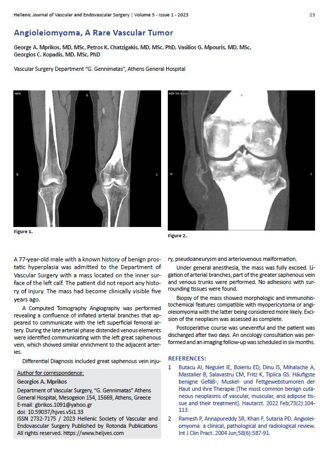
*A 77-year-old male with a known history of benign prostatic hyperplasia was admitted to the Department of Vascular Surgery with a mass located on the inner surface of the left calf. The patient did not report any history of injury. The mass had become clinically visible five years ago. A Computed Tomography Angiography was performed revealing a confluence of inflated arterial branches that appeared to communicate with the left superficial femoral artery. During the late arterial phase distended venous elements were identified communicating with the left great saphenous vein, which showed similar enrichment to the adjacent arteries.
Differential Diagnosis included great saphenous vein injury, pseudoaneurysm and arteriovenous malformation. Under general anesthesia, the mass was fully excised. Ligation of arterial branches, part of the greater saphenous vein and venous trunks were performed. Νo adhesions with surrounding tissues were found. Biopsy of the mass showed morphologic and immunohistochemical features compatible with myopericytoma or angioleiomyoma with the latter being considered more likely. Excision of the neoplasm was assessed as complete. Postoperative course was uneventful and the patient was discharged after two days. Αn oncology consultation was performed and an imaging follow-up was scheduled in six months
References
- Butacu AI, Negulet IE, Boieriu ED, Dinu IS, Mihalache A, Mastalier B, Salavastru CM, Fritz K, Tiplica GS. Häufigste benigne Gefäß-, Muskel- und Fettgewebstumoren der Haut und ihre Therapie [The most common benign cutaneous neoplasms of vascular, muscular, and adipose tissue and their treatment]. Hautarzt. 2022 Feb;73(2):104-113.
- Ramesh P, Annapureddy SR, Khan F, Sutaria PD. Angioleiomyoma: a clinical, pathological and radiological review. Int J Clin Pract. 2004 Jun;58(6):587-91.

