A Huge Ruptured Pseudoaneurysm of the Radial Artery Presented One Year after the Coronary Intervention – A Case Report
| Available Online: | July, 2026 |
| Page: | 79–81 |
Author for correspondence:
George Galyfos
email: georgegalyfos@hotmail.com
Address: 114 Vasilissis Sofias Avenue, Athens, Greece, 11527
Tel.: +30 2132088132
Fax: +30 2107707574
ORCID ID: 0000-0002-6077-007X
Doi 10.59037/4qrrqz53
Stavros Patsouris, Panagiotis Theodorou, Nikolaos Intzes, Anna Mexi, Konstantinos Saliaris, Andreas Theodorou, Argyris Athanasiou, Kitsou Eleni, George Galyfos
Vascular Surgery Unit, First Department of Propaedeutic Surgery, National and Kapodistrian University of Athens, Hippocration Hospital, Athens, Greece.
Full Text
Gallery
Full Text
Abstract
Introduction: Radial artery pseudoaneurysms (PSAs) are rare, especially when they are of huge size and present with rupture. There is no consensus regarding optimal management, and treatment varies according to size and presentation.
Case Report: We aim to present a case with a late rupture of a huge radial artery PSA, one year after coronary intervention.
Conclusion: Early detection and treatment is always recommended when the PSA increases in size or shows signs of imminent rupture.
Keywords: radial artery, pseudoaneurysm, rupture, skin necrosis, large size, treatment
INTRODUCTION
Radial artery pseudoaneurysms (PSAs) presenting after coronary interventions are extremely rare, with an estimated incidence of <0.1% according to literature.1 They are usually associated with pain and discomfort if they keep increasing in size. If they are left untreated, they could lead to expansion and rupture.2 Ruptured radial artery PSAs are even rarer, and they could threaten the patient’s life if not treated emergently.1
We aim to report on the case of a huge radial artery PSA presented with a late rupture and treated in our department.
CASE REPORT
A 74-year-old patient with a history of coronary intervention and stent placement one year ago, atrial fibrillation (AF), and depression was transferred to our emergency department (ER) with active bleeding due to a huge radial artery PSA on his right forearm. The patient was under per os antiplatelet and anticoagulant treatment for both his coronary disease and AF. Although his personal cardiologist had diagnosed the PSA many months ago and suggested that the patient should visit a vascular surgeon, the patient did not follow his advice which led to the expansion and rupture of the PSA.
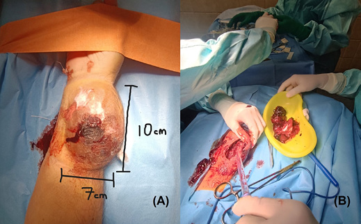
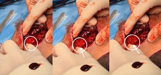
The PSA and the point of bleeding were securely pressed with bandages in the ER, and the patient was transferred immediately to the operating room (OR). The patient was still hemodynamically stable despite the blood loss although serum hemoglobin levels were lower than normal. The PSA was 10 x 7 cm in size with central skin necrosis due to expansion (Fig. 1A). Under general anesthesia and proximal arterial occlusion with an external tourniquet device, a longitudinal incision was made and all the necrotic skin was removed. A large amount of thrombus and necrotic pseudo-membranes were removed (Fig. 1B) and the point of rupture was detected after depressurizing the tourniquet (Fig. 2). The artery was sutured with non-continuous Prolene 6-0 sutures. Additionally, hemostatic dressings were placed within the wound cavity due to diffuse bleeding (possibly due to the combined antiplatelet and anticoagulant treatment).
The radial artery was palpable distally after the closure of the artery. A vacuum drainage was placed and the skin was closed with Nylon 3-0 sutures. The postoperative course of the patient was uneventful.
Discussion
Both a radial artery PSA delayed presentation and rupture are extremely rare in literature. Additionally, there is no consensus regarding proper management. In a large retrospective study by Harvey et al. including over 10,000 radial catheterizations, the incidence of a radial artery PSA was 0.06%.3 Radial artery PSAs present after different types of interventions such as arterial line placement or coronary artery catheterization. They present usually after removal of the catheter or the sheath and inadequate pressure appliance over the puncture site.4 The artery is mostly affected at the distal part where its position is more superficial. A significant predisposing factor is anticoagulant or dual antiplatelet treatment.4 Also, local infection can be a predisposing factor.5 According to literature, most of the cases progress slowly and only one third of the cases will require an urgent operation.1
The patient may present early after the intervention with local swelling or a pulsatile mass, pain, or discomfort.6 Local signs of inflammation or infection may be present. Although the PSA could be asymptomatic when it is small, it may be painful when it increases in size. The ultrasound remains the first-choice method to diagnose the PSA and to determine its morphology as well. If it increases in size or when a local infection is present, the risk of rupture is higher. However, in some cases, a computed tomographic angiography (CTA) may be necessary.1,4
When an intervention is required, several treatments have been suggested. A percutaneous thrombin injection under ultrasound guidance remains one option.7 However, one should keep in mind that a wide PSA neck will increase the risk for failure of PSA occlusion and for distal thrombin embolism. Additionally, the PSA could be occluded right after the thrombin injection and a recurrence could be detected during follow-up, especially if the patient is under full anticoagulant treatment.7 Another option is plain ultrasound guided compression (UGC).8 However, thrombin injection has been found to be superior to UGC as it achieves thrombosis within 6 seconds compared to UGC that may require up to 45 minutes.8 UGC can also cause rupture to larger PSAs as well. According to some authors, UGC may be selected for pediatric cases.8 When the PSA is under 2 cm in diameter, there is a 70% possibility that it will spontaneously get thrombosed. Spontaneous thrombosis may occur within 4 weeks. UGC may have an effectiveness between 60% and 90%.8
However, infection, skin ischemia and frail appearance of the PSA are contraindications for any pressure appliance.9 In these cases, an open repair is recommended. Our patient presented late after the intervention (almost a year) with a huge, ruptured PSA combined with skin necrosis, that needed emergency surgical treatment. Usually, the point of arterial rupture is found and a simple suturing of the artery is adequate such as in our case. However, ligation of the artery remains an alternative when anastomosis or suturing the artery is not possible.9 Ligation should be performed when it is tested that the ulnar artery is patent, when there are good collaterals in the angiography or excellent back bleeding from the distal stump.6 Finally, there are some case reports referring to endovascular treatment of radial artery PSAs either with covered stent placement or embolization. However, these cases are extremely rare.10
CONCLUSION
In conclusion, ruptured artery PSAs of large size are rare but life-threatening. Therefore, early detection and treatment is always recommended when the PSA increases in size or shows signs of imminent rupture. When ruptured, open repair is the most frequent strategy with the aim to preserve the distal perfusion.
REFERENCES
- Dey S, Baruah A, Mohanta PK. Ruptured pseudoaneurysm of the radial artery. BMJ Case Rep. 2009;2009:bcr06.2008.0080.
- Filis K, Galyfos G, Sigala F, Karantzikos G, Vavouranakis M, Toutouzas K, Albanopoulos K, Zografos G. Vascular complications during transcatheter aortic valve implantation: The role of the vascular surgeon. Vascular. 2020;28:421-429.
- Harvey JA, Kim S, Ireson ME, Gulati R, Bell MR, Moran SL. Acute Upper-Limb Complications Following Radial Artery Catheterization for Coronary Angiography. J Hand Surg Am. 2020;45:655.
- Galyfos G, Kerasidis S, Stefanidis G, Stamatatos I, Kastrisios G, Giannakakis S, Geropapas G, Papacharalampous G, Maltezos C. Iatrogenic and non-iatrogenic arterial injuries in an urban level I trauma center in Greece. Int Angiol. 2016;35:526-30.
- Ganchi PA, Wilhelmi BJ, Fujita K, Lee WP. Ruptured pseudoaneurysm complicating an infected radial artery catheter: case report and review of the literature. Ann Plast Surg. 2001;46:647-50.
- Galyfos G, Chamzin A, Charalampopoulos G, Alexakis N, Sigala F, Filis K. Emergent management of life-threatening iatrogenic femoral artery injuries – Report of four cases. HJVES. 2023;3:142-147.
- Reus M, Vázquez V, Alonso J, Morales D, Rodríguez JM. Treatment of a radial artery pseudoaneurysm with ultrasound-guided percutaneous thrombin injection in a patient with Behçet’s syndrome. J Clin Ultrasound. 2003;31:440-444.
- Molina-López V, Nieves-La Cruz C, Llopart-Herrera L, Mirabal-Arroyo J. Successful treatment of radial artery pseudoaneurysm after transradial cardiac catheterization with continuous compression therapy by a TR Band® radial compression device. Cardiovasc Revasc Med. 2021;28: 227-231.
- Papadoulas SI, Tsantrizos P, Kouri N, Tsimpoukis A, Koutsogiannis N, Moulakakis KG, Kakkos SK, Davlouros P. A case report of surgical repair of a post-catheterization radial pseudoaneurysm. Pan Afr Med J. 2022;41:261.
- Tsiafoutis I, Zografos T, Koutouzis M, Katsivas A. Distal radial access for percutaneous endovascular repair of a radial artery pseudoaneurysm. J Invasive Cardiol. 2020;32:198.