Open conversion for atypical endoleak after EVAR without endograft explantation: a case report
| Available Online: | July, 2026 |
| Page: | 74-78 |
Author for correspondence:
Dr. Serena Arianna Cutolo
Division of Vascular Surgery, IRCCS San Raffaele Scientific Institute, Vita-Salute San Raffaele University, Via Olgettina 60, 20132, Milan, Italy
email: cutolo.serenaarianna@hsr.it
Tel: +39 02-26437130 Fax: +39 02-26437148
Doi 10.59037/e37f8121
Serena Arianna Cutolo, * Kerolos R. Basta, * Carlotta Bugna, Fiorenza De Lisio, Germano Melissano
*The first two authors have contributed equally to the writing of the manuscript
Division of Vascular Surgery, IRCCS San Raffaele Scientific Institute, Vita-Salute San Raffaele University, Milan, Italy
Full Text
Gallery
Full Text
Abstract
Introduction: Endoleaks are significant complications following Endovascular Aortic Aneurysm Repair (EVAR). However, persistent aneurysm sac enlargement without an identifiable endoleak remains challenging.
Case Report: We report a 78-year-old man who developed progressive aneurysm sac expansion to 64 mm five years after EVAR despite prior inferior mesenteric artery embolization. Computed Tomography Angiography (CTA) demonstrated focal delayed enhancement within the aneurysm sac without evidence of type I, II, or III endoleak. Open surgical exploration revealed diffuse oozing from the intimal surface without identifiable branch inflow or endograft defect. Sacotomy with endograft preservation was performed, including oversewing of a small bleeding vessel, adjunctive argon plasma coagulation (APC), and fibrin sealant application. The postoperative course was uneventful, and follow-up imaging demonstrated sac shrinkage without recurrent endoleak.
Conclusion: This case highlights open surgical repair with endograft preservation as a valuable option for persistent sac enlargement after EVAR when endovascular treatment fails or no discrete bleeding source is identified in selected patients.
Keywords: EVAR, atypical type II endoleak, aneurysm sac expansion, open conversion, endograft preservation
INTRODUCTION
With the development of endovascular aortic techniques, endovascular aortic aneurysm repair (EVAR) has become a primary approach for the treatment of Abdominal Aortic Aneurysms (AAA).1 However, aneurysm sac enlargement not responsive to endovascular treatment represents a leading cause of reintervention after EVAR.2 Persistent sac expansion can lead to potential rupture, which compromises not only the endograft but also the patient’s life.3,4
Current clinical guidance emphasizes imaging-based characterization of post-EVAR sac enlargement, prioritizing the exclusion of type I and III ELs before attributing sac growth to type II EL, endotension, or hidden sources.5 The European guidelines define atypical type II EL (aT2EL) as sac expansion in the absence of a clearly defined endoleak cavity or identifiable arterial inflow, often associated with delayed-phase mural enhancement and hypertrophied adventitial vasa vasorum on imaging; this entity has also been described as “Moyamoya endoleak” in selected reports.6,7
This report describes a case of persistent aneurysm sac enlargement after EVAR despite prior embolization of the Inferior Mesenteric Artery (IMA), ultimately managed with open sacotomy and endograft preservation. Written informed consent for publication was obtained from the patient.
CASE REPORT
A 78-year-old man was referred to our center because of progressive aneurysm sac enlargement reaching 64 mm, five years after EVAR performed with a Medtronic Endurant stent graft (Medtronic, Minneapolis, MN, USA). The aneurysm sac had progressively enlarged despite previous embolization of the inferior mesenteric artery performed six months before presentation.
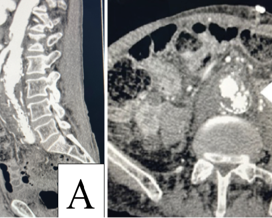
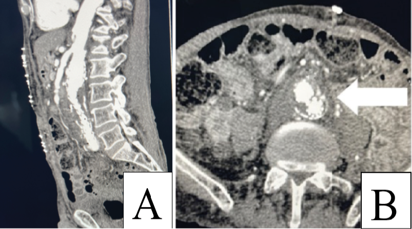
Computed tomography angiography (CTA) performed at admission demonstrated the absence of patent lumbar arteries or inferior mesenteric artery inflow, with focal delayed enhancement within the aneurysm sac thrombus. No evidence of type I, II, or III endoleak was identified. The endograft remained patent, with preserved perfusion of the iliac vessels (Figure 1Α, 1Β).
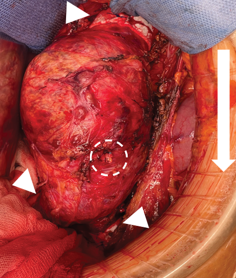
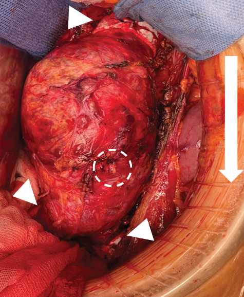
On presentation, the patient was asymptomatic and hemodynamically stable. His medical history was significant for end-stage renal disease requiring chronic hemodialysis. Open surgical repair was performed through a xipho-umbilical midline laparotomy using an Alexis® retractor (Applied Medical Resources Corp., Rancho Santa Margarita, CA, USA) (Figure 2). The aneurysm was exposed from the level of the renal arteries to the iliac bifurcation. The aneurysm sac appeared tense but non-pulsatile, and intra-sac pressure measurements confirmed the absence of pulsatility. The proximal aortic neck and iliac arteries were encircled with Teflon felts and secured using transfixing 2-0 polypropylene sutures incorporating both the endograft and the native aortic wall. The previously embolized IMA was identified, dissected, transected, and ligated using 4-0 polypropylene sutures (Figure 2).
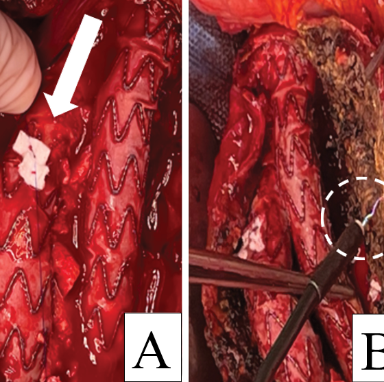
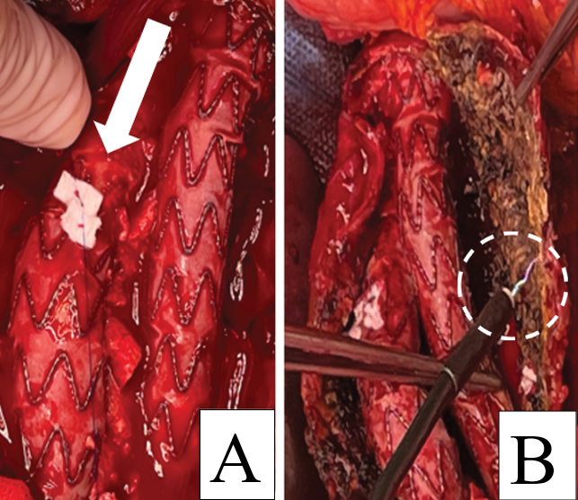
The aneurysm sac was opened without aortic cross-clamping. Following decompression, the cavity contained chronically organized thrombus mixed with fresh blood. After thrombus evacuation, diffuse oozing from the aneurysm sac intimal surface was observed without identification of a discrete bleeding source. The proximal and distal endograft attachment sites appeared intact. Additional transgraft fixation sutures were placed at the overlap of the iliac limb to improve mechanical stability and prevent device displacement during surgical manipulation (Figure 3).
A small posterior wall bleeder was oversewn to achieve focal hemostasis. Because diffuse oozing persisted, coagulation diathermy and Argon plasma coagulation (APC; Erbe Elektromedizin GmbH, Tübingen, Germany) were applied to the aneurysm sac surface to achieve complete hemostasis (Figure 3Α, 3Β).
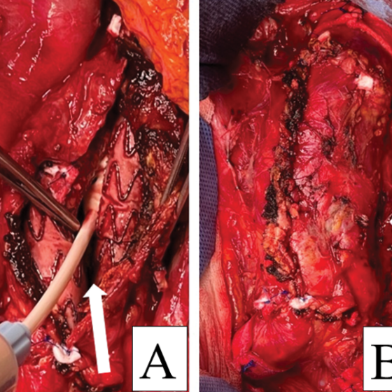
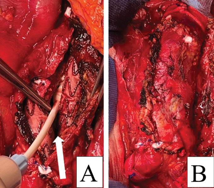
After sac reduction, adjunctive fibrin sealant was applied to reinforce hemostasis (Figure 4). The residual aneurysm sac was then closed over the preserved endograft, and the retroperitoneum was drained and closed (Figure 4Α, 4Β).
The postoperative course was uneventful, and the patient was discharged on postoperative day 4. The CTA performed at three months demonstrated sustained reduction in sac size, with no evidence of EL.
DISCUSSION
Endoleaks remain one of the principal determinants of long-term EVAR durability. Persistent aneurysm sac enlargement in the absence of a clearly identifiable EL represents a particularly challenging clinical scenario because occult sac pressurization may ultimately result in aneurysm rupture. Although the exact criteria requiring intervention have not been definitively established, continued sac growth despite repeated imaging surveillance generally warrants active treatment.8
Several methods for managing type II ELs have been reported. To prevent aneurysm sac enlargement after EVAR, perioperative coil embolization via trans-arterial or trans-lumbar approaches is the most common method. Despite the success of this technique in treating type II ELs, repeated interventions are frequently required. Notably, 51% of patients treated with coil embolization alone undergo additional procedures. High rates of subsequent embolizations are reported in those undergoing these procedures. These reports suggest that patients require regular surveillance10 after coil embolization and may require multiple interventions. Repeated interventions carry additional risks, including infection, nephrotoxicity from contrast agents – particularly relevant in patients with chronic renal failure – and increased radiation exposure during CT-guided trans-lumbar procedures.3,4
Current European Society for Vascular Surgery (ESVS) guidelines recommend consideration of elective open conversion, with or without endograft preservation, in patients with persistent aneurysm sac enlargement despite prior endovascular treatment attempts.5 These recommendations underscore the role of open surgical repair as a valuable therapeutic option after failure of less invasive strategies.
Nevertheless, open surgical repair remains technically demanding and more invasive than embolization, with potentially increased morbidity and mortality related to aortic cross-clamping, endograft-associated inflammatory changes, and anastomotic bleeding.7 The prevalence of advanced age, cardiovascular disease, pulmonary comorbidities, and impaired physiological reserve among EVAR patients further contributes to perioperative risk.
Sacotomy with preservation of the endograft offers several potential advantages because it avoids complete graft explantation and may eliminate the need for aortic cross-clamping. This approach may therefore reduce operative stress while allowing direct treatment of sac pressurization, and being feasible also in high – risk patients. However, sacotomy also has limitations, including the possibility of missed occult ELs, incomplete treatment, and future aneurysm sac re-expansion. Consequently, long-term postoperative imaging surveillance remains mandatory.
An interesting case report published by Kinugasa et al.8 showed that one of the potential causes of EL is a microvascular conduit from the adventitia to the lumen across an injured intima-media interface. They reached this conclusion after treating a patient with persistent EL 10 years after EVAR, despite successful embolization of the IMA. Histopathological assessment of the sac showed a distinctive triad of: extensive intimal denudation, media exposure with luminally oriented microvessels, and adventitial vasa vasorum proliferation.
Onitsuka et al.9 demonstrated that endoaneurysmorrhaphy that combines sacotomy, ligation of the back-bleeding vessels, and endograft preservation for type II EL can reduce operation time and result in a less invasive OSR than with graft removal. Short-term outcomes included shrinkage of the aneurysm sac with a stable diameter, no missed type II EL, no type II EL recurrence, and no evidence of endograft migration or disjunction. In addition, in cases of endograft preservation, the proximal neck banding method may further prevent aneurysm re-enlargement and type Ia EL secondary to neck-dilatation.
Ultimately, with the development of endovascular techniques, there is a gradual trend toward replacing open surgery. However, in complex cases, such as those with complicated anatomy or EVAR repair failure, traditional embolization materials or catheter techniques may not fully exclude ELs. In these cases, operative time may be prolonged, radiation exposure may increase, and the risk of surgical failure is higher. Additionally, repeated endovascular repairs may lead to long-term complications such as aneurysm sac enlargement, graft wear, infections, and other issues, which may sometimes require open surgery for definitive resolution.1
CONCLUSION
Management of atypical type II endoleaks (aT2ELs) associated with persistent aneurysm sac enlargement after EVAR remains challenging, particularly after failed embolization attempts. In selected patients, open surgical repair with endograft preservation represents a valuable treatment option when further endovascular or minimally invasive approaches are not feasible or have proven ineffective. This strategy allows direct control of sac pressurization, prevents further aneurysm sac enlargement, and may contribute to the long-term durability of the previous EVAR repair while avoiding the morbidity associated with complete graft explantation.
BIBLIOGRAPHY
- Li Z, Chen Z, Cai J, Liu Z, Li X, Qiao T. Outcomes of Open Surgical Repair for Post-EVAR Endoleaks: A Retrospective Cohort Study with Mid-term Follow-Up. Ann Vasc Surg. 2026 Jan. doi:10.1016/j.avsg.2025.12.033 PubMed PMID: 41534712.
- Iwakoshi S, Ogawa Y, Dake MD, Ono Y, Higashihara H, Ikoma A, et al. Outcomes of embolization procedures for type II endoleaks following endovascular abdominal aortic repair. J Vasc Surg. 2023 Jan;77(1):114-121.e2. doi:10.1016/j.jvs.2022.07.168
- Chaikof EL, Dalman RL, Eskandari MK, Jackson BM, Lee WA, Mansour MA, et al. The Society for Vascular Surgery practice guidelines on the care of patients with an abdominal aortic aneurysm. J Vasc Surg. 2018 Jan 1;67(1):2-77.e2. doi:10.1016/j.jvs.2017.10.044 PubMed PMID: 29268916.
- Chun JY, de Haan M, Maleux G, Osman A, Cannavale A, Morgan R. CIRSE Standards of Practice on Management of Endoleaks Following Endovascular Aneurysm Repair. Cardiovasc Intervent Radiol. 2024 Feb 1;47(2):161-76. doi:10.1007/s00270-023-03629-1 PubMed PMID: 38216742.
- Wanhainen A, Van Herzeele I, Bastos Goncalves F, Bellmunt Montoya S, Berard X, Boyle JR, et al. Editor’s Choice – European Society for Vascular Surgery (ESVS) 2024 Clinical Practice Guidelines on the Management of Abdominal Aorto-Iliac Artery Aneurysms. European Journal of Vascular and Endovascular Surgery. 2024 Feb 1;67(2):192-331. doi:10.1016/j.ejvs.2023.11.002 PubMed PMID: 38307694.
- Torikai H, Inoue M, Nakatsuka S, Tamura M, Yashiro H, Yoshitake A, et al. Imaging Findings of Atypical Type II Endoleak Through Vasa Vasorum After Abdominal Endovascular Aneurysm Repair. Cardiovasc Intervent Radiol. 2018 Jan 31;41(1):186-90. doi:10.1007/s00270-017-1778-y
- Yamada M, Takahashi H, Tauchi Y, Satoh H, Matsuda H. Open Surgical Repair Can Be One Option for the Treatment of Persistent Type II Endoleak after EVAR. Ann Vasc Dis. 2015;8(3):210-4. doi:10.3400/avd.oa.14-00133
- Kinugasa Y, Tai R, Teshima H. Wall Derived Atypical Type II Endoleak 10 Years after Endovascular Aneurysm Repair: Intra-operative and Histopathological Evidence of Vasa Vasorum Mediated Haemorrhage. EJVES Vasc Forum. 2026 Feb. doi:10.1016/j.ejvsvf.2026.02.003
- Onitsuka S, Ito H. Surgical Treatment of Sac Enlargement Due to Type II Endoleaks Following Endovascular Aneurysm Repair. Ann Vasc Dis. 2023 Mar 25;16(1):1-7. doi:10.3400/avd.ra.22-00115
- Sarac TP, Gibbons C, Vargas L, Liu J, Srivastava S, Bena J, et al. Long-term follow-up of type II endoleak embolization reveals the need for close surveillance. J Vasc Surg. 2012 Jan;55(1):33-40. doi:10.1016/j.jvs.2011.07.092