Emergency management of life-threatening iatrogenic femoral artery injuries – report of four cases
| Available Online: | October, 2023 |
| Page: | 142-147 |
Author for correspondence:
George Galyfos
Vascular Surgery Unit, First Department of Propedeutic Surgery, National and Kapodistrian University of Athens, Hippocration Hospital, 114 Vasilissis Sofias Avenue, 11527, Athens, Greece
Τel: +30 2132088132
Fax: +30 2107707574
E-mail: georgegalyfos@hotmail.com
doi: 10.59037/ye0vdr55
ISSN 2732-7175 / 2023 Hellenic Society of Vascular and Endovascular Surgery Published by Rotonda Publications All rights reserved. https://www.heljves.com
Abstract
Full Text
References
Images
Abstract
Abstract:
Iatrogenic vascular injuries (IVIs) have increased in the last decades due to the increase in endovascular interventions. The common femoral artery (CFA) is a very common location of IVI. Although many cases are treated conservatively or with minimally invasive techniques, management of life threatening IVIs may be quite challenging. This report aims to present four uncommon cases with life-threatening iatrogenic CFA injuries that were treated with open surgery and discuss on the proper management.
Full Text
INTRODUCTION
Iatrogenic vascular injuries (IVIs) have increased in number during the last decades, with an estimated incidence of more than one third of all vascular trauma cases.1 Due to the increase of endovascular procedures for the treatment of coronary artery disease (CAD) and peripheral artery disease (PAD), percutaneous arterial puncture and sheath introduction has become a very common practice.2 There are several options considering the access vessel for performing such endovascular procedures although the common femoral artery (CFA) remains very popular, especially for more complex procedures.3 The occurrence of iatrogenic complications after CFA puncture including haemorrhage, pseudoaneurysm (PSA) formation or acute thrombosis depends on several factors such as length of operation, type of the sheath, antithrombotic treatment, obesity, performance of puncture under ultrasonographic guidance or not, adequate compression, the presence of PAD and others.2,4
When the injury is small or stable, more conservative treatments such as compression or ultrasound-guided thrombin injection (UGTI) have been applied with success.5 However, when the injury is life-threatening and the patient becomes unstable, an emergency repair is indicated.2 In many cases, establishing control of the bleeding and repairing the injury may be challenging, especially when the hematoma is of massive size. This report aims to present four unusual cases with a life-threatening IVI post catheterization of the CFA, and discuss on proper management.
CASE 1
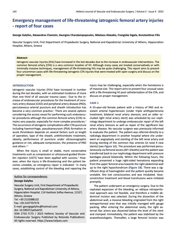
A 55-year-old female patient with a history of PAD and resistant arterial hypertension (under triple antihypertensive treatment; bilateral renal artery stenosis; and an older occluded right renal artery stent) was scheduled by our nephrology department to undergo endovascular repair of the left renal artery stenosis as well as repair of the significant iliac artery disease. No vascular surgeon was previously informed to evaluate the patient. The patient was referred directly to a radiology department in another hospital where she underwent an angioplasty and stenting of the left renal artery and kissing stenting of the common iliac arteries (in total 4 new stents) (see Figure 1A). The procedure was performed percutaneously via femoral access (6Fr sheaths) and the patient was transferred back to our nephrology department with pressure bandages placed bilaterally. Within the following hours, the patient presented a huge right-sided hematoma expanding from the upper femoral area towards the right abdominal wall up to the right hypochondrium (Figure 1B). There was a significant drop of haemoglobin and the patient quickly became unstable. She lost consciousness and was intubated. Vasoconstriction treatment and blood transfusions were initiated promptly.
The patient underwent an emergency surgery. Due to the cephalad expansion of the bleeding, an oblique retroperitoneal approach was not feasible, and therefore a middle abdominal incision was selected. During the dissection of the abdominal wall, a massive bleeding originated from the right extraperitoneal area that was initially managed with gauge packing. After entering the abdominal cavity, the right external iliac artery was dissected below the level of the stent and clamped. Immediately, the patient was stabilized by the anaesthesiologists. Thereafter, a large femoral incision was made, and the femoral bifurcation was dissected. Two rupture points were identified in the CFA and sutured with Prolen 5-0. (Figure 1C) All wounds were closed, and drains were placed both under and over the inguinal ligament. The patient was extubated and transferred to intensive care unit (ICU) for close monitoring. The patient was discharged a week after in optimal condition.
CASE 2
A 52-year-old female patient with obesity (BMI>30kg/m2), PAD and chronic renal disease (under peritoneal dialysis) underwent a scheduled open mechanical mitral valve replacement due to severe regurgitation in our cardiac surgery department. Intraoperatively, a triple-lumen 8Fr arterial sheath was placed into the left CFA to monitor arterial pressure. Postoperatively, the sheath was removed under coumarin treatment for the valve (INR > 2.5). Possibly due to full anticoagulation and suboptimal pressure appliance, a huge expanding hematoma presented in the upper left femoral area that led to significant haemoglobin drop, severe pain and incipient skin necrosis (Figure 2A). The patient underwent a computed angiography (CTA) illustrating a 17cmX14cm hematoma and an extravasation from the left CFA (Figure 2B). Before being transferred to operation room, the patient started becoming hemodynamically unstable and vasoconstriction treatement was initiated. As there was no expansion of the hematoma above the inguinal ligament, an oblique retroperitoneal approach was selected for proximal control. A second incision was made below the hematoma and superficial femoral artery was also dissected for distal control. Then, a third incision was made over the hematoma. A large amount of blood as well as thrombus were drained. During dissection of the femoral vessels, a longitudinal rupture (almost 1.5cm in length) of the common femoral vein (CFV) was identified that was sutured with Prolen 5-0. At the same level, a small rupture site of the CFA was also detected that was also sutured with Prolen 5-0. (Figure 2C) All wounds were closed, and 4 drains were placed overall. The patient was extubated and transferred to ICU for close monitoring. She was discharged after 5 days from the hospital in optimal condition.
CASE 3
A 58-year-old male patient with a history of CAD underwent a scheduled percutaneous coronary intervention (PCI) in the interventional cardiology department of our institution. The procedure was conducted via a left CFA access and pressure bandages were placed after the removal of the sheath. However, the patient within the following hours presented an large expanding hematoma of the left femoral area, with the femoral circumference being almost twice the size of the contralateral limb. (Figure 3) The patient quickly became hemodynamically unstable, and he received 4 blood units due to significant haemoglobin drop. Without delay, the patient was transferred to the operating room and underwent emergency surgery. Due to the size of the hematoma, an oblique extraperitoneal approach was again selected for proximal control. After controlling the external iliac artery, a femoral incision was made. Again, two rupture points were identified, one in the left CFV and one in the left CFA. Both ruptures were sutured with Prolen 5-0. The patient was stabilized and transferred to ICU for close monitoring. He was discharged within a week in optimal condition.
CASE 4
A 72-year-old patient with history of cardiac valve disease and obesity was scheduled for a transcatheter aortic valve implantation (TAVI). The procedure was conducted percutaneously via a left CFA access in the interventional cardiology department of our institution. After the removal of the sheath (16Fr), the cardiologists placed a percutaneous closing device twice without success. The patient quickly became unstable due to haemorrhage through the puncture site. A cut-down dissection was performed immediately within the angio-suite and clamps were placed in the left CFA to stop the bleeding. (Figure 4A) The patient then was transferred to the operating room (in another floor) and underwent an emergency surgery. The femoral bifurcation was dissected, and a severe injury of the entire CFA was detected. Therefore, the CFA was substituted with an interposition 7mm PTFE graft. (Figure 4B) The patient was stabilized and transferred to ICU for close monitoring. There were no signs of ischemia in the limb. The patient was discharged after one week in optimal condition.
DISCUSSION
Arterial interventions are associated with an overall minor complication rate of less than 10%, and major complications requiring transfusion or surgical intervention occur at a rate of less than 1%.4 However, injury of the femoral artery is a complication with an incidence reaching up to 3.5% after transfemoral procedures in interventional cardiology and haemodialysis units.6 Most arterial injuries present as a pulsatile mass, and they may have a palpable thrill or an audible murmur. The diagnosis is confirmed with ultrasound imaging as first-line option.1,4 The ultrasound can identify the point of extravasation, the amount of thrombus, the blood flow velocity pattern and all the associations with adjacent structures. However, a further angiographic imaging may be necessary for the following reasons: a) when ultrasound is inconclusive; b) there are signs of ischemia; c) the injury is extended to the retroperitoneal area; d) an endovascular repair is planned.7
When the patient becomes unstable like in our cases, the indication for emergency repair is set without the need for diagnostic imaging.4 An injury of the CFA can cause major bleeding, PSA formation, thrombosis or distal embolization, local pain, neuropathy or local skin necrosis.6 Risk factors include large-sized catheters, false technique, obesity, haemodialysis, hypertension, anticoagulation and PAD, concurring with our cases.6 Patients treated for CAD or valve disease usually receive large doses of antiplatelets or anticoagulants that increase the risk for bleeding as well. Furthermore, patients with chronic renal disease may have also some degree of coagulopathy.7
Life-threatening IVIs of the femoral artery are rare in general, but they can be devastating.6 In a recent study evaluating life-threatening IVIs, however, groin was the most common location (42%).8 Considering each specialty, IVIs were detected in the groin in 66% of interventional radiology cases and in 33% of thoracic surgery cases, concurring with our report.8 Additionally, transfemoral access is associated with higher mortality compared to transradial access for endovascular repair of PAD or carotid disease.9 It seems that death if occurs, it is not attributable to the injury itself in all cases. However, almost half of the injuries are considered avoidable with possible causes including false communication, doctor’s delay, technical false, false patient selection or indication.8,10 In our cases, the use of ultrasonographic guidance, a more careful pressure appliance, or the use of a closing device could have prevented major bleeding in some of the patients. In addition, a vascular surgeon was not informed preoperatively in all the cases and that may have impacted the outcome significantly. We have found in an earlier study that a standardized preoperative evaluation by a vascular surgeon was associated with a lower mortality risk among patients undergoing TAVI.10 Even after the injury, delay in obtaining vascular surgery assistance may lead to unnecessary blood loss or additional vascular damage.9
Non-surgical treatment remains the primary management for femoral IVIs when the patients are asymptomatic and the diameter of the PSA is < 2cm. Indications for open surgery include hemodynamic instability of the patient, rapid expansion of a hematoma/PSA, mycotic infection of a PSA, compromised soft tissue viability, failure of more conservative strategies, concomitant distal ischemia and neurological deficit due to local pressure.1,2,5 Conservative management may be also preferred in patients with many comorbidities and significant anaesthetic risk. This management must include restoration of the patient’s haemostatic function, lowering blood pressure, possible transfusions and absolute bed rest. Of course, frequent haemoglobin measurements and close clinical assessment are essential so that the plan may be changed promptly to surgical treatment if necessary.1,2,5 The success rate of conservative treatment is significantly lower compared to surgery although the complications rate is higher after surgical repair.5 When comparing conservative strategies, it seems that UGTI has superior results compared to ultrasound-guided compression.5 UGTI may be associated with rare complications such as peripheral embolization, anaphylactic reaction to thrombin and skin infection. Some data indicate that sac area and neck length/width is associated with the success of UGTI.5
When intervention is indicated, open surgery is usually reserved for rupture or expanding PSAs. Although endovascular treatments (covered stent placement or coil embolization) are currently used in many cases, their efficacy and safety have been shown only in small studies.5 Almost half of femoral IVIs are treated with simple suture of the vessel like in our cases.7 When the damage is more extent, a more complex repair may be needed such as end-to-end anastomosis, patch placement, bypass or interposition grafting. Vascular injuries have a better prognosis when treated quickly after injury. In cases of large hematomas with possible retroperitoneal expansion, a proximal external iliac artery dissection and control should be used whereas in cases with obvious bleeding below the inguinal ligament, dissection of the CFA only may be adequate for controlling the bleeding. In some cases, a concomitant venous injury may be present that can cause major bleeding despite the proximal arterial control concurring with our report.4 In these cases, the attempt to obtain vascular control with forceful use of clamps may result in additional injuries compared to achieving haemostasis with compression. Digital compression or the use of swabs may be a more appropriate strategy for venous injuries.
In conclusion, the majority of IVIs in the femoral area are avoidable and a standardized preoperative evaluation by a vascular surgeon may prevent them. They should be treated with open repair promptly when they are expanding, ruptured or complicated. Especially in cases of rapid retroperitoneal expansion of a hematoma or giant-sized PSAs, a proximal control above the inguinal ligament should limit the blood loss. Caution should be taken as a concomitant major venous injury may be present and increase the amount of blood lost.
References
- Filis K, Sigala F, Stamatina T, Georgia D, Zografos G, Galyfos G. Iatrogenic Vascular Injuries of the Abdomen and Pelvis: The Experience at a Hellenic University Hospital. Vasc Endovascular Surg. 2019;53:541-546.
- Filis K, Arhontovasilis F, Theodorou D, Albanopoulos K, Lagoudianakis E, Manouras A, Vavuranakis M, Vlachopoulos C, Toutouzas K, Tsiamis E, Androulakis A, Kallikazaros I, Giannopoulos A, Bramis I, Stefanadis C. Management of early and late detected vascular complications following femoral arterial puncture for cardiac catheterization. Hellenic J Cardiol. 2007;48:134-42.
- DeRubertis BG, Faries PL, McKinsey JF, Chaer RA, Pierce M, Karwowski J, et al. Shifting paradigms in the treatment of lower extremity vascular disease: a report of 1000 percutaneous interventions. Ann Surg. 2007;246:415-24..
- 4. Ge BH, Copelan A, Scola D, Watts MM. Iatrogenic percutaneous vascular injuries: clinical presentation, imaging, and management. Semin Intervent Radiol. 2015;32:108-22.
- Wu H, Zhang L, Zhang C, Xie B, Lou C, Liu Y, Bai H. Non-Surgical treatment Versus Surgery for Iatrogenic Femoral Artery Pseudoaneurysms: Systematic Review and Meta-Analysis. Front Surg. 2022;9:905701.
- Kulkarni LM, Sirsat SM. Ruptured femoral artery pseudoaneurysm: A life-threatening, iatrogenic catastrophe! Indian J Anaesth. 2016;60:437-9.
- Rammos C, Rassaf T. Insight in the treatment of iatrogenic femoral artery pseudoaneurysms. Vasa. 2021;50:163-164.
- Rudström H, Bergqvist D, Björck M. Iatrogenic vascular injuries with lethal outcome. World J Surg. 2013;37:1981-7.
- Galyfos G, Sigala F, Filis K. Transradial versus Transfemoral access in patients undergoing peripheral artery angioplasty/stenting: A meta-analysis. Cardiovasc Revasc Med. 2018;19:457-465.
- Filis K, Galyfos G, Sigala F, Karantzikos G, Vavouranakis M, Toutouzas K, Albanopoulos K, Zografos G. Vascular complications during transcatheter aortic valve implantation: The role of the vascular surgeon. Vascular. 2020;28:421-429.


